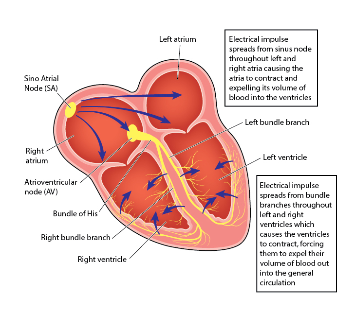
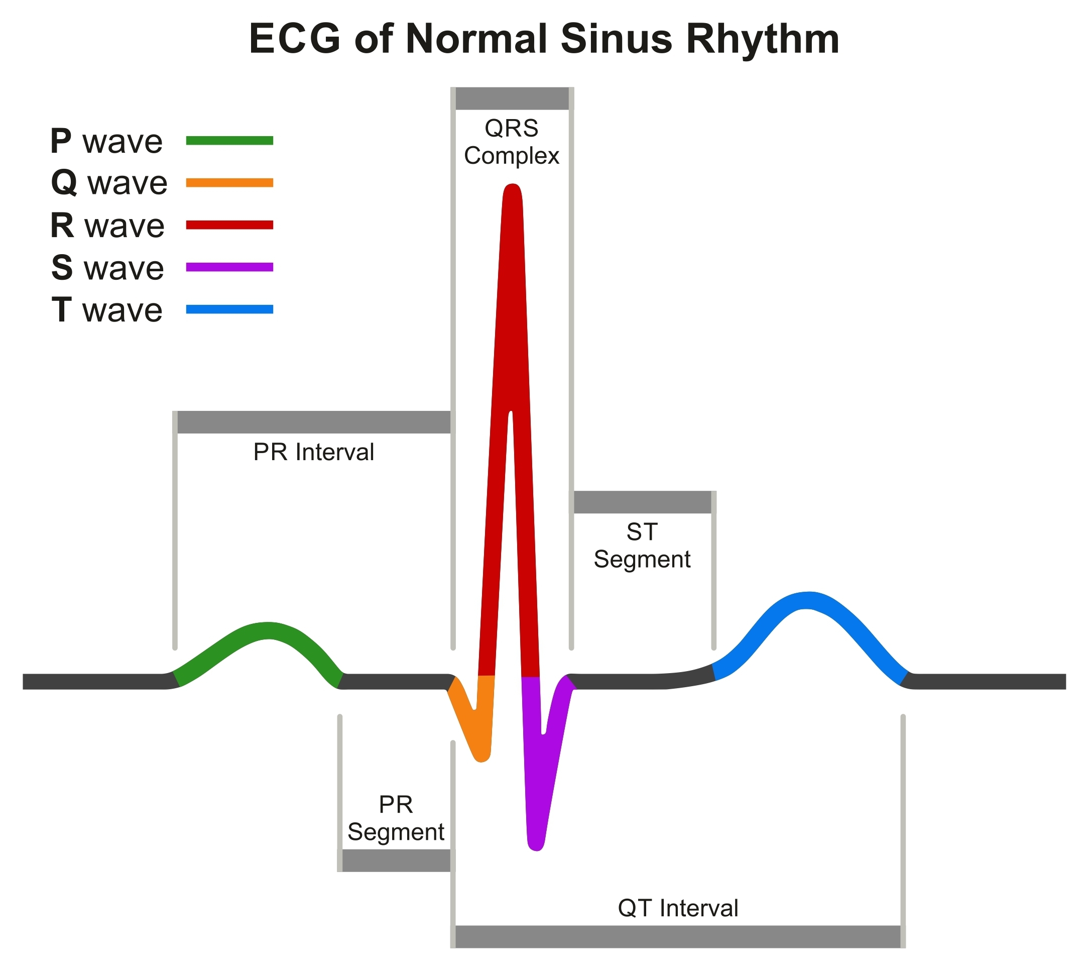
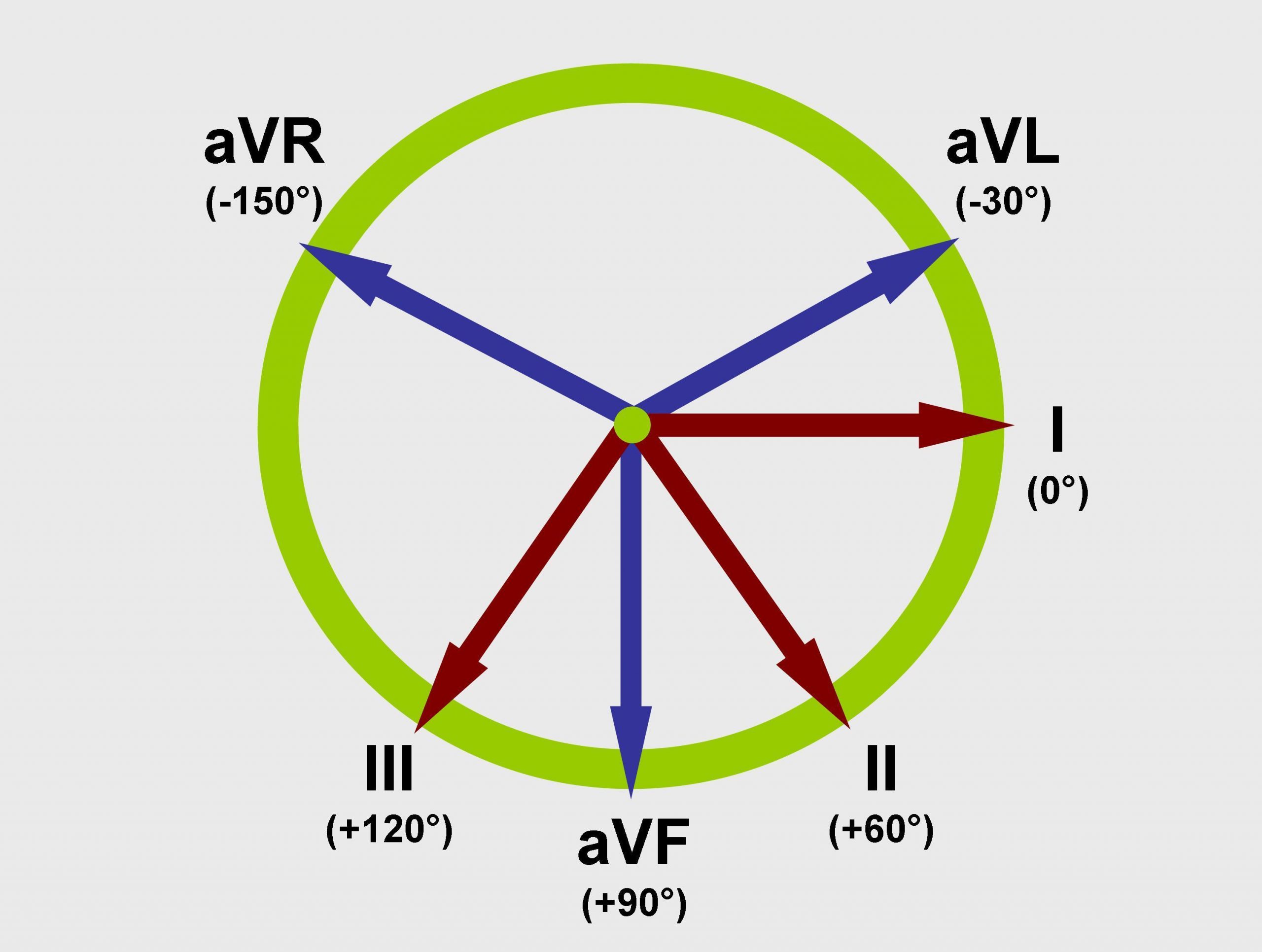
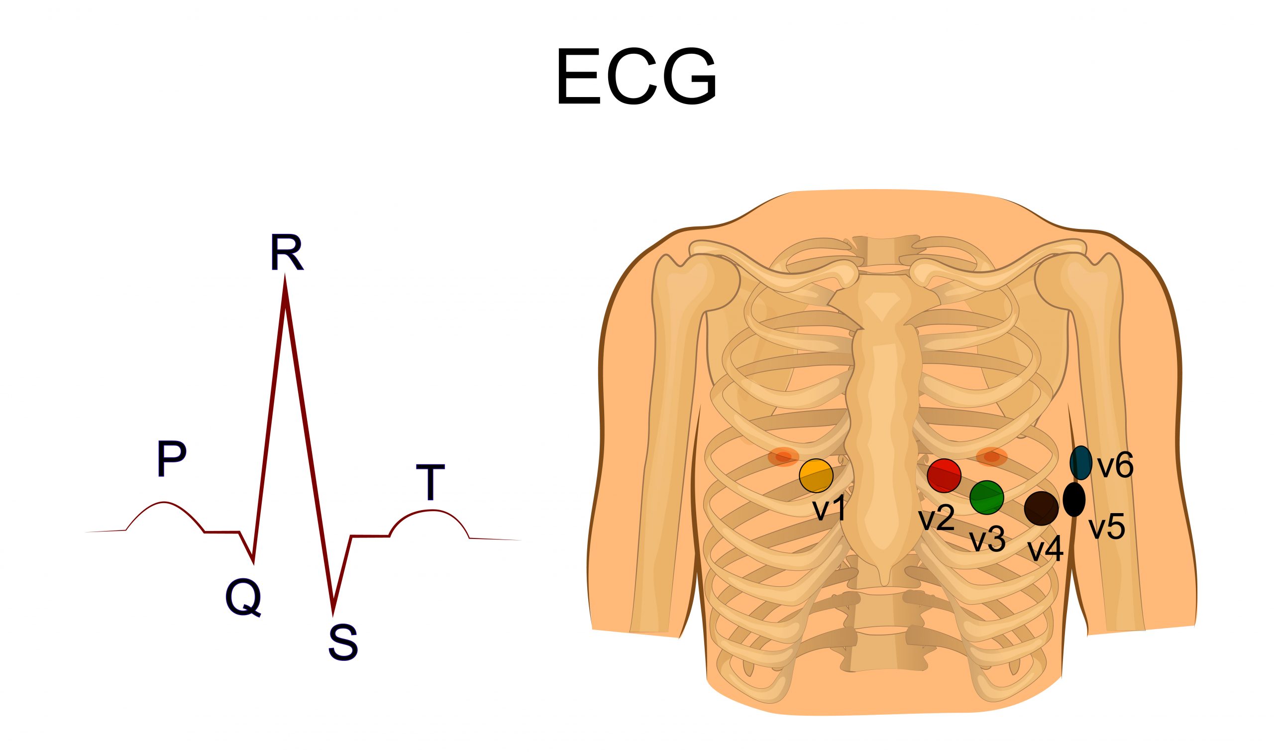
Basics of EKG Approach to EKGTermClinical featuresSinoatrial (SA) node 1 mm cell collection in upper right atrium with intrinsic depolarization rate at 60-100 beats/minInitiates atrial systole and generates P waveAtrioventricular (AV) nodeLocated near inferior portion of interatrial septumProvides delay for atria to send blood to ventricles before initiating ventricular systoleProtects ventricles from excessive stimulation from atriaPR interval indicates how well AV node is functioningHis bundles-Purkinje systemLeft bundleDivides into left anterior and posterior fascicles that differentiate into Purkinje fibers.Supplied by left anterior descending (LAD) arteryRight bundleInnervates right ventricle to depolarize myocardiumBundles and Purkinje fibers allow for smooth depolarization of ventricles and correspond to QRS complexWaveforms P wave Indicates impulse traveled from SA node to AV nodeSymbolizes atrial depolarizationQRS complex Indicates ventricular depolarizationImpulse has traveled from atria through AV node and into His bundles systemT wave Indicates ventricular repolarizationImpulse has traveled ventricles and back to atriaU wave Delayed repolarization abnormalityUsually seen in hypokalemiaVector of impulseImpulse traveling towards a lead creates upward (positive) deflectionImpulse traveling away from lead creates downward (negative) deflectionMeasurementsPaper speed Typically at 25 mm/secSmall boxes1 mm representing 40 msecLarge boxes5 small boxes (5 mm) representing 0.2 secVertical boxesEvery 10 mm represents 0.1 mV of electrical potentialLead placement Limb leads (I, II, III, aVF, aVR, and aVL) Look at heart from frontal planePrecordial leads (V1-V6) Look at heart from vertical planeLesson Intro Video https://www.dropbox.com/scl/fi/i8e9xyjzy2crogj5qzi8f/Approach-to-Acid-Base-Part-1-Physiology-Review.mp4?rlkey=jnuar31h8ht9kvygra5xubevr&dl=0(Next Lesson) Rate Back to Approach to EKG No Comments Comments are closed.
 Atrioventricular (AV) nodeHis bundles-Purkinje systemAtrioventricular (AV) nodeHis bundles-Purkinje system

No Comments